
QUALITY EYECARE FOR YOUR ENTIRE FAMILY
Standard Eye Exam
A complete eye examination involves a series of tests designed to evaluate your vision and check for eye diseases. Optometrists use a wide variety of tests and procedures, which range from simple ones, like having you read an eye chart, to complex tests, such as using a high-powered lens to visualise the tiny structures inside your eyes.
A comprehensive eye examination can take an hour or more, depending on the number and complexity of tests required to fully evaluate your vision and the health of your eyes. The optometrist will usually do the following:
- Eye tests to determine your refractive error and prescription. Instruments such as an autorefractor or retinoscope may also be used.
- Cover tests that check how well your eyes work together.
- Eye movement testing (ocular motility), to assess how well your eyes follow a moving object.
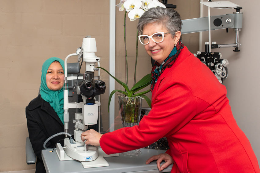
- Slit-lamp examinations that give your optometrist a magnified view of the structures in your eyes in order to determine their health.
- Ophthalmoscopy which allows the optometrist to evaluate the back of the eye and detect ocular disease. Early signs of certain systemic diseases (e.g.diabetes and high blood pressure) may be seen on the retina before they are diagnosed medically.
- Sometimes your optometrist may decide to take a ‘base line’ photograph of the back of your eye, using a fundus camera.
- Eye pressure or “air puff” tests that check for glaucoma.
In some cases, besides these common tests performed during a standard comprehensive eye examination, your optometrist may recommend other, more specialised eye tests.
Diagnosis of Eye Diseases
Many eye diseases have no early symptoms. They may be painless, and you may see no change in your vision until the disease has become quite advanced.
The single best way to protect your vision is through regular professional eye examinations.
Of course, between examinations, if you notice a change in your vision – or you think your eye may be injured in any way – contact your eye care professional immediately.
Comrehensive Eye Health Exam
Many people aren’t sure what to expect when they make an appointment with an optometrist especially if they’ve never had a comprehensive eye exam before or it’s been many years since their last exam.
Optometrists use a wide variety of tests and procedures to examine your eyes. These tests range from simple ones, like having you read eye chart, to complex tests, such as using a high-powered lens to visualize the tiny structures inside of your eyes.
Eyes are sensitive indicators and may reveal vision problems, eye diseases and general health irregularities long before any obvious physical symptoms
exist. Health care professionals recommend that all people should have periodic and thorough eye tests as part of their routine primary health care.
expect the following assessment
Analysing all your current eyewear – Distance only and or readers only/ Progressive lenses / Office lenses and prescription sunglasses. Your current prescription will be assessed according to your current prescription of each lens including the optical needs required.
Your optometrist will now have a complete overview of your sight and vision abilities and will explain and will discuss recommendations. You will be introduced to one of our optical dispensers who will assist you with taking measurements to align the centre of the optical lens with centre point of eye and take in consideration all your natural the facial features.
The Zone of Clear Single Binocular vision describes the range of accommodation (focus) alongside the range of vergence (eye teaming) to give a picture of the full range within which a patent achieves clear, single vision and comfort.
If your comprehensive eye examination reveals any medical condition requiring further examination, the optometrist will refer you to your family doctor, or an ophthalmologist.
Step 1: Health and Medication History Assessment
As part of your vision history, your Camarena Porter optometrist may ask you the following questions:
- Your overall health and that of your immediate family.
- The medications you are taking (prescription and over-the-counter).
- Questions about high blood pressure (hypertension), diabetes, smoking, and sun exposure.
- How well you can see at present, including any recent changes in your vision.
- Eye diseases that you or your family members have had, including macular degeneration and glaucoma.
- Previous eye treatments, surgeries, or injuries.
- The date of your last eye examination and age of current prescription.
- Are you having any problems with your vision?
- Do you have any allergies?
- Finally, your optometrist will ask you the main reason for an eye test and visual requirements for distance and near vision and all symptoms.
- Sensitive to glare day and or night
- Hobbies that optical needs
- Safety optical needs to protect from hazards
Your own health and that of your family will give the optometrist an indication of any issues that may be affecting, or could affect, your vision
Step 2: Visual Acuity (measure your ability to see)
Step 2.1: Retinoscopy
Your optometrist may perform this test early in the eye exam to obtain an approximation of your optical lens prescription to obtain best sight (visual acuity).
In retinoscopy, the room lights will be dimmed and you will be asked to focus on a large target (usually the big "C" on the eye chart). As you stare at the "C," your optometrist will shine a light at your eye and flip lenses in a machine (phoropter) in front of your eyes. This test estimates which lens powers will best correct your distance vision.
Step 2.2: Refraction
This is the test technique the optometrist uses again with phoropter / trail optical lenses in trail frame, to determine and refine the final optical correction for distance and near.
During a refraction, the optometrist puts the instrument called a phoropter in front of your eyes and shows you a series of lens choices. The optometrist will then ask you which of the two lenses in each choice looks clearer.
Based on your answers, your optometrist will continue to fine-tune the lens power until reaching a final eyeglass prescription.
Step 2.3: Autorefractor
Your optometrist also may use an autorefractor to automatically estimate your optical lens prescription. With both devices, a chin rest stabilizes your head while you look into the instrument at a pinpoint of light or a detailed image.
An autorefractor, like a manual refraction, determines the lens power required to accurately focus light on your macula on the retina.
The autorefraction takes only a few seconds, and the results greatly reduce the time required for your optometrist to perform a manual refraction and determine your optical correction to correct myopia, hyperopia, astigmatism and presbyopia.
Step 3: Eye Muscle Assessment
Your Camarena Porter optometrist evaluate your eye muscle balance to make sure your eyes are co-ordinated and working together in all directions at distance and near The images formed by each eye are correctly aligned to have clear comfortable single vision.
The cover test is the simplest and most common technique to evaluate the “alignment posture” of the eyes and prism bars the “stamina of alignment” between the 2 eyes.
During these tests, your eye doctor will assess whether the uncovered eye must move to pick up the fixation target, which could indicate strabismus (or other problem that could cause eye strain or amblyopia (“lazy eye”).
3.1 Ocular motility testing (eye movements)
Ocular motility testing is performed to determine how well your eyes can follow a moving object and/or quickly move between and accurately re- fixate on two separate targets in all 9 positions of gaze.
Testing of smooth eye movements ("pursuits") is more common. The optometrist will ask head to follow the slow movement of a hand-held light or other target with just your eyes.
If quick eye movements (“saccades”) also are tested, your optometrist will ask to focus between two targets positioned some distance apart from each other. The optometrist could also use the DEM test to analyse.
Problems with eye movements can cause eye strain and may affect reading ability on paper / digital screens and skills on sport field or driving behind a steering wheel.
3.2 Stereopsis test (depth perception)
Stereopsis is the term used to describe eye teaming that enables normal depth perception and appreciation of the 3-dimensional nature of objects.
In one commonly used stereopsis test, you wear a pair of “;3D” glasses and look at a booklet of test patterns.
3.3 Accommodation (focus ability)
MEM retinoscopy and accommodative test will evaluate the “posture” of your accommodative system and the flipper test the stamina and flexibility of your “focus stamina”.
There is a relationship between your ability to focus near and your age and the period of sustained bear vision. The near add will be determined.
Recommendations to maintain clear focused vision for required working distances will be discussed with you and the recommended lens design.
3.4 Dominant eye
Determine the dominant eye and dominant hand, that need to be noted for reading and writing. Dominant eye also important, in some calculations of fitting contact lenses.
Step 4: Interior & Exterior Eye Examination
Your optometrist will carefully examine the exterior and interior of your eyes whilst conducting the following assessments:
- Ophthalmoscopy- Detailed interior eye examination, which can give an overall picture of your eyes and your general health. Various parts of the eye such as the lens, the vitreous and the fundus (includes retina and optic nerve) are assessed.
- Field of Vision Test – asses the width of visual field and to locate your normal blind spot.
- Slit Lamp Test – used to screen the eye lids, lashes and front segment of the eye. Fluorescein strips and cobalt light to evaluate health of cornea and
lacrimal system in contact lens patients and tear break up time. - Tonometry Test – air puff, that gentle measure the internal eye pressure of the eye, as one of the screening tests for glaucoma.
- Colour Vision Test – used to evaluate your ability to perceive colours.
- Keratometry Test – measures the curvatures of the front curvature of the eye and also used for contact lens fitting
- Digital imaging of the fundus – photo of the interior layer of the optic nerve and blood supply and light sensitive cells at the macula
- Lacrimal evaluation – evaluation of the tear’s quality and quantity
- Amsler grid assist to detect early signs of retinal disease and monitor changes in vision